In 1886, Weil published detailed case descriptions of 4 patients with icterus, splenic enlargement and nephritis [1]. All four patients presented with acute onset of fever, elevated liver function tests (LFTs), hepatic enlargement, splenomegaly, and proteinuria. Two cases were recorded more than12 years earlier, but the author felt that they shared significant similarities with 2 more recent cases from 1882. Initially, patients presented with a high fever accompanied by general malaise, headaches, somnolence and even delirium. Many had GI symptoms. He mentions that iridocyclitis was noticeable in one patient and a roseola-like skin rash in another.
The overall course of the illness was benign – there were no fatalities – and patients did not develop hepatic failure despite highly abnormal LFTs and marked jaundice. Of note, in 3 patients, the disease was diphasic with milder symptoms reappearing after a symptom free interval Treatment was attempted with methods typical for the era and consisted of calomel (mercury chloride), quinine, muriatic acid and such, but nothing seemed to do any good.

Weil had no biopsy reports, no cultures, and not even microscopic demonstration of a potential pathogen. Nonetheless, the author felt strongly that the disease must be infectious in nature. He goes over the differential diagnosis in great detail, yellow fever, recurrent fever, acute hepatitis, typhus including ‘formes frustes’. These are discussed and dismissed.
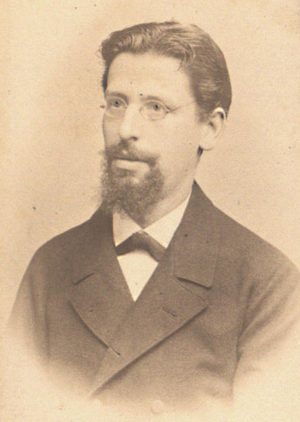
Weil certainly displayed extraordinary clinical acumen. He made connections and drew conclusions based on the knowledge of only 4 cases, occurring many years apart, recognizing a pattern of signs and symptoms that he felt were due to a yet unknown infectious etiology. While he was convinced that this was a new clinical entity, he remained cautious pointing out the lack of supporting evidence from pathology or bacteriology.
He had an extraordinary almost prophetic sense of foresight and proved to be correct, but it took another 30 years to confirm his prescient assumptions.
Actually, the discovery of the pathogen causing Weil’s disease is yet another amazing story. The spirochete was identified during the first world war, around the year 1915. Visualized with special stains and observed on darkfield microscopy, a new spirochete, leptospira, was identified as the cause of Weil’s disease by several research groups almost simultaneously, first in Japan and a few months later in Germany. Their efforts went on in parallel and took place independently. In the span of only 12 months, a series of publications appeared, mutually confirming the organism’s size and shape, propagation in guinea pigs, short presence in blood but high density in the liver, asymptomatic carrier state in rodent kidneys. With communication hampered by the war in Europe, these research groups were not aware of one another, and it was not clear in the beginning who had first identified the Weil bacillus.

Indeed, a 1916 article in the New York Times [2] wrongly attributed the discovery of the spirochete to Uhlenhuth who had published a detailed description of his experimental work in the March 1916 issue of the Berliner Klinische Wochenschrift.
Uhlenhuth was already a prominent scientist in those days and contributed much to early leptospirosis research. He was unable to infect cats, dogs, pigs, monkeys or rats; only guinea pigs and hamsters came down with the disease after inoculation with infectious blood. The ‘virus’ could be passaged over several generations in guinea pigs. He described spirochetes with peculiar endings in liver specimens using Levanditi staining, a treponemal silver nitrate-based method and saw spirochetal movement on darkfield microscopy. He found organisms in large numbers in liver cells, but rarely in blood. When immunity developed, it was transferable and partially protective. He even tested various chemotherapeutic agents, including Atoxyl and Neo-Salvarsan, arsenicals used for the treatment of syphilis and trypanosomiasis.
As it turned out, Uhlenhuth’s publication was preceded by the work of another group of German researchers who published their findings in a series of ‘announcements’. Hübener and Reiter were in hot pursuit of a spirochete, which they saw on liver stains as mentioned in their first of 4 reports from 1915. However, their claim to have found the etiology of Weil’s disease drew criticism from Dr. Weil himself who felt that the identified pathogen may not be causative of Weil’s disease as he had originally described it. Hübener’s detailed response provided more data in support of the spirochetal origin and seemed to have settled the discourse.
Hübener and his team in Berlin had indeed identified a spirochete, but – unbeknownst to them and Uhlenhuth (and the NYT) – Japanese researchers can claim priority by a few months. It was Inada and coworkers who first announced the discovery of a leptospiral pathogen in early 1915. They published their findings on the spirochete which they called Leptospira icterohaemorrhagicae. We refer you to Dr. Kobayashi’s account who in 2001 chronicled the timeline of leptospirosis research ultimately leading to the discovery of the pathogen.[1]
The race to the finish line involved at least 3 research groups, all converging on the final identification of the pathogen we now call L. interrogans. All contributed to our knowledge as far as epidemiology and diagnosis are concerned.
A timeline of key events, articles and publication references is provided in the Table below (the Japanese references according to Kobayashi [1]):
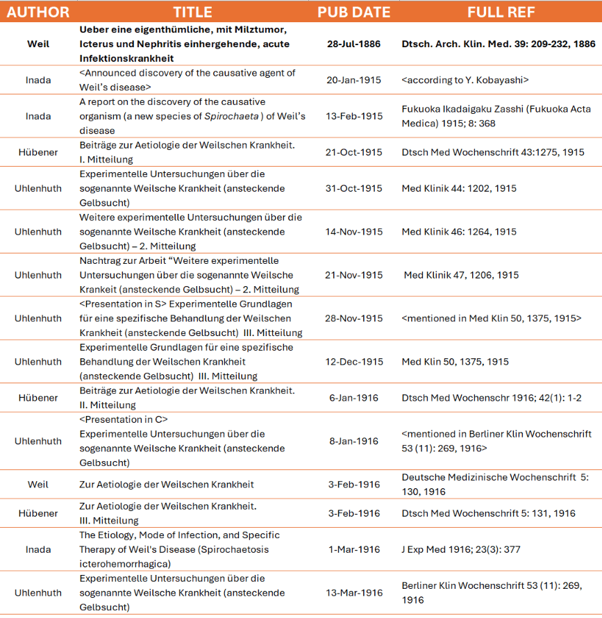
It becomes clear that keeping track of the rapid fire of publications must have been difficult for reasons of language, publication in lesser known journals, geographic distance and war-related communication issues.
The history of leptospirosis is fascinating. It features a clinician with an uncanny ability for recall of patient histories who reports a new disease. He is proven right by the concerted efforts of microbiologists who – 30 years later – in quick succession were able to identify the pathogen of Weil’s disease. Rightly so, Inada, Uhlenhuth and Weil are still recognized for their contributions to medicine, serology and bacteriology.
The story does not end here. There are several cross-connections that are probably less known, like the debate about animal experimentation, defended by Uhlenhuth, the training of Japanese researchers in German labs including Inada, the role of news media sensationalizing claims that prove only partially correct, and later career and role of some key investigators during the Third Reich (Uhlenhuth, Reiter).
There is an important zoonotic component to leptospirosis. While uncommon in humans, uveitis is fairly common in horses. Also called ERU (equine recurrent uveitis) or ‘moon blindness’, it is an immune phenomenon secondary to leptospirosis frequently leading to blindness. Antibiotics are not helpful; treatment is with steroids and NSAIDs.
The underlying immune pathology of leptospira has been getting morre attenttion in recent years. Associations with genetic markers, interleukins like IL-6, TNF, IFNγ, and pentraxins are under investigation.
Existing diagnostic tests for leptospirosis seem antiquated. The tedious microscopic agglutination test (MAT) and other serologic assays to detect a titer increase between early and convalescent sera, ELISA and PCR tests are still mentioned in textbooks; they have significant shortcomings which result in underreporting of the disease. A recent article takes a new approach by studying virulence factors.[3] There is clearly a need for more specific and less labor-intensive tests that could be brought to countries with limited resources. Making a diagnosis of leptospirosis by bacteriology is just as difficult today as it was for Inada, Uhlenhuth, and Huebener.
PS1. The Weil-Felix reaction for rickettsial typhus is named after Edmund Weil.
No relationship to Adolf Weil of leptospirosis fame!
PS2. Adolf Weil is sometimes listed as the discoverer of the atypical amino acid norleucine.[4] This is incorrect: Norleucine was discovered by Arthur Weil in 1913 together with the somewhat controversial biochemist Emil Abderhalden (who had a minor asteroid named after him).[5],[6]
REFERENCES (See also Table above)
[1] Kobayashi Y. Discovery of the causative organism of Weil’s disease: historical view. J Infect Chemother 7:10, 2001
[2] New York Times: Find the Cause of Weil’s disease – Oct 1, 1916
[3] Chaurasia R. Early Diagnosis of Human Leptospirosis by Detection of Antibodies to Leptospira-Secreted Virulence-Modifying Protein Exotoxins. Am. J. Trop. Med. Hyg., 114: 17, 2026
[4] Adolf Weil (physician) – Wikipedia (accessed 9/12/2026)
[5] Abderhalden E. Spaltung von dl-Aminocapronsäure (= Norleucin) in die optisch-aktiven Komponenten mittels der Formylverbindung. Polypeptide, an deren Aufbau Aminocapronsäure beteiligt ist. Hoppe Seyler’s Zschrift f. Physiol. Chem. 86: 454, 1913
[6] Emil Abderhalden u. Arthur Weil. Vergleichende Untersuchungen über den Gehalt der verschiedenen Bestandteile des Nervensystems an Aminosäuren. 2. Mitteilung Hoppe Seyler’s Zschrift f. Physiol. Chem. 83: 425, 1913
